➤DISSECTION ➛Cut through the muscles arising from the lateral and medial epicondyles of humerus and reflect them distally if not already done.
Also cut through biceps brachii, brachialis and triceps brachii 3 cm proximal to the elbow joint and reflect them distally. Remove all the muscles fused with the fibrous capsule of the elbow joint and define its attachments.
Features
The elbow joint is a hinge variety of synovial joint between the lower end of humerus and the upper ends of radius and ulna bones.
Elbow joint is the term used for humeroradial and humeroulnar joints. The term elbow complex also includes the superior radioulnar joint also.
➤Articular Surfaces
Upper
The capitulum and trochlea of the humerus. The coronoid fossa lies just above the trochlea and is designed in a manner that the coronoid process of ulna fits into it in extreme flexion. Similarly the radial fossa just above the capitulum allows for radial head fitting in the radial fossa in extreme flexion.
Lower
i. Upper surface of the head of the radius articulates with the capitulum.
ii. Trochlear notch of the ulna articulates with the trochlea of the humerus . The elbow joint is continuous with the superior radioulnar joint. The humeroradial, the humeroulnar and the superior radioulnar joints are together known as cubital articulations.
Ligaments
1. Capsular ligament:➛ Superiorly , it is attached to the lower end of thehumerus in such a way thatthe capitulum, the trochlea, the radial fossa, the coronoid fossa and the olecranon fossa are intracap srtlar. lnfer omedially, it is attached to the margin of the trochlear notch of the ulrra except laterally; inferolaterally , it is attached to the annular ligament of the superior radioulnar joint. The synovial membrane lines the capsule and the fossae, named above. The anterior ligament, and the posterior ligament are thickening of the capsule.
2 The ulnar collateral ligament is triangular in shape ➛. Its apex is attached to the medial epicondyle of the humerus, and its base to the ulna. The ligament has thick anterior and posterior bands: These are attached below to the coronoid process and the olecranon process respectively. Their lower ends are joined to each other by an oblique band which gives attachment to the thinner intermediate fibres of the ligament. The ligament is crossed by the ulnar nerve and it gives origin to the flexor digitorum superficialis. It is closely related to the flexor carpi ulnaris and the triceps brachii
3. The radial collateral or lateral ligament:➛ It is a fan-shaped band extending from the lateral epicondyle to the annular ligament. It gives origin to the supinator and to the extensor carpi radialis brevis .
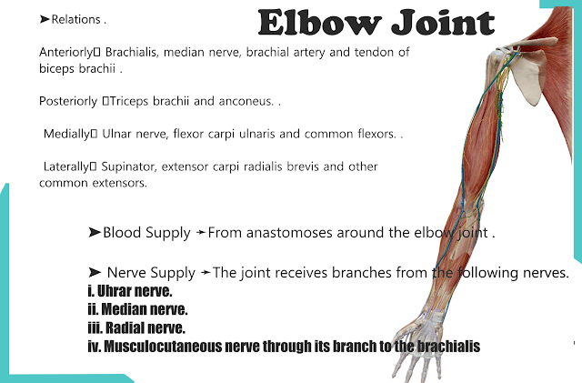
➤Relations .
Anteriorly➛ Brachialis, median nerve, brachial artery and tendon of biceps brachii .
Posteriorly ➛Triceps brachii and anconeus. .
Medially➛ Ulnar nerve, flexor carpi ulnaris and common flexors. .
Laterally➛ Supinator, extensor carpi radialis brevis and other common extensors.
➤Blood Supply ➛From anastomoses around the elbow joint .
➤ Nerve Supply ➛The joint receives branches from the following nerves.
i. Uhrar nerve.
ii. Median nerve.
iii. Radial nerve.
iv. Musculocutaneous nerve through its branch to the brachialis.
➛ Movemenls
1 Flexion is brought about by:
i. Brachialis.
ii. Biceps brachii.
iii. Brachioradialis.
2 Extension is produced by:
i. Triceps brachii.
ii. Anconeus.


Post a Comment
If you have any doubts any queries so contact me on social media account and send mail